国家天元数学西北中心“图像质量评价和图像质量评价引导的CT重建专题研讨班”定于12月13日-15日在西安交通大学举办。该研讨班为中心2019“数据科学与医疗健康”主题年活动之一。本期专题研讨班将聚焦医学图像质量评价模型和方法、CT重建成像方法、图像多层次结构与图像质量的关系等热点问题,邀请相关专家“以半天一位专家”的形式介绍专题成果,与研讨班学员交流并研讨最新研究进展。
一、专题研讨班日程安排
时间:2019年12月13日-15日 上午8:30-11:30 下午14:30-17:30
地点:西安交通大学数学楼二楼2-1会议室
活动召集人:牟轩沁教授 西安交通大学

二、招生对象:
计划招生50人左右。全国高等院校和科研机构从事医学、人工智能、应用数学及相关交叉领域的青年教师及在校博士生均可报名参加。
三、学员待遇
专题研讨班免收培训费,学习期间为全体学员提供餐补。根据中心促进西北地区学科发展,活动向西部地区倾斜的原则,研讨班为新疆、青海、宁夏、甘肃地区学员安排住宿。
四、报名参与方式
有意参加的青年教师及博士生请于12月1日前将附件报名信息(见附件)及个人简历发送到:xbty@xjtu.edu.cn。邮件标题请注明“申请人姓名+单位+12月专题研讨班报名”。国家天元数学西北中心组织委员会将对申请人材料进行审定,并于12月6日前邮件通知入选者本人。如未接到录取通知即为未入选,不再另行通知。
五、联系方式
联系人:白老师
办公电话:029-82665627
邮箱:xbty@xjtu.edu.cn
地址:西安交通大学数学与统计学院111办公室
六、课程介绍
活动召集人:牟轩沁 西安交通大学
牟轩沁,博士,教授。任西安交通大学图像处理与识别研究所所长、国家数据广播工程技术研究中心主任。担任国家自然科学基金委员会第十二届学科专家评议组成员,中国图像图形学会常务理事,中国体视学会常务理事,中国体视学会CT专业委员会副主任委员,陕西省图像图形学会副理事长。担任过多个国内外期刊的编委会成员和国际学术会议的程序委员会成员职务。2017年组织召开了CT/PET/SPECT研究、开发和应用领域权威的国际会议(两年一次):The 14th Fully Three-Dimensional Image Reconstruction in Radiology and Nuclear Medicine (Fully3D2017),并担任大会主席。主要研究方向是成像技术和计算机视觉,主持了近20项纵向基金项目,其中包括国家“863计划”重大项目和国家重点研发计划课题;获得过教育部新世纪优秀人才支持计划和骨干教师基金的资助;在科研成果方面,与合作单位共同开发了国内最早的CCD型医用X线电视系统;主持完成的项目“RA3900II型数字减影系统”获得了教育部发明类科技进步二等奖(1999年,第一获奖人);发表了经同行评议的学术论文200余篇(期刊论文和会议论文各100余篇),发表论文的SCI引用总数3600余次, Google Scholar引用总数6500余次,其中包括4篇ESI论文;获得国家发明专利授权10余项。
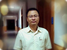
课程题目1:Bayesian Image Reconstruction and Bayesian Classifier
主讲人:Jerome Zheng Liang, Professor, Stony Brook University
专家简介:
Jerome Zheng Liang gained Ph.D. degree in Physics from City University of New York in 1987, followed by one year Research Fellow in Nuclear Medicine and Radiation Oncology at Albert Einstein College of Medicine. He had been a Research Associate and Assistant Professor in Radiology at Duke University Medical Center. He joined State University of New York at Stony Brook (SBU) in 1992 and currently holds professorship in Departments of Radiology, Electrical and Computer Engineering, Computer Science, and Biomedical Engineering. He was a co-founder of the Program in Biomedical Engineering at SBU. His primary research interests in medical imaging include data acquisition geometry, image formation and processing methodology, and feature-based visualization and computer-aided detection and diagnosis. He has authored more than 400 scientific publications, 20 US patents, 45 invited talks and 170 conference presentations. He has supervised more than 25 postdoctors and 40 graduate students (PhD and MS degrees). He has served principal investigator (PI) for 11 NIH projects and Co-PI for four NIH projects. He received the State University of New York Chancellor's Entrepreneur Award for “whose invention has led to the startup of a company to commercialize the product” in 2002. He was elected Fellow to the IEEE for “contributions to medical image reconstruction and virtual colonoscopy” in 2007. He received the 1st “Excellence in Translational Research” Award in SBU School of Medicine (2013).
授课内容:
Medical diagnosis is one of the most fundamental tasks for healthcare. Great research efforts have been devoted to explore new diagnostic methods and improve existing methods. Medical imaging methods have been playing an essential role in medical diagnosis, because of their non-invasiveness. Two important components in medical imaging methods are reconstructing the acquired raw data for the human body images and classifying the reconstructed images for decision- or diagnostic making.
Quantifying the image reconstruction performance is necessary to judge if the explored new methods have the feasibility to advance the diagnostic field, and if the proposed improvement of an existing method has a significant gain.
Similarly, quantifying the image classification performance is also necessary to judge if the explored new methods have the feasibility to advance the diagnostic field, and if the proposed improvement of an existing method has a significant gain.
This lecture will take low-dose computed tomography (CT) image reconstruction and computer-aided diagnosis of polyps from reconstructed CT colonography images.
课程题目2:图像质量评价中的问题、理论与方法
主讲人:张林 副教授 同济大学
专家简介:
张林博士于2003年和2006年在上海交通大学获得学士和硕士学位。之后曾先后供职于Microsoft和Autodesk公司。2008年3月至香港理工大学攻读博士学位,导师为张磊教授。2011年8月加入同济大学。主要研究兴趣为机器视觉与图像理解。他以第一或通信作者身份已在IEEE T-PAMI、IEEE T-IP、IEEE T-MM、Pattern Recognition、ACM MM等期刊和会议上发表论文60余篇。根据Google Scholar统计,其所发表论文总的被引用次数为5841次;其中,4篇论文入选ESI Highly Cited Papers。其论文“FSIM: A feature similarity index for image quality assessment, IEEE Trans. Image Processing, 20 (8) 2378-2386, 2011”为IEEE TIP自2011年以来所有发表论文中被引用次数最高的论文,目前被引用2353次。他于2013年入选上海市浦江人才计划。他目前是IEEE高级会员、中国计算机学会(CCF)高级会员、中国计算机学会(CCF)计算机视觉专业委员会委员、视觉与学习青年研讨会(VALSE)资深领域主席(Senior Area Chair)。
授课内容:
基于图像的应用系统在我们的生活中几乎已经无处不在。而图像质量评价是图像获取与分析领域中的一个重要而基本的问题,它会影响到我们对元器件的选型及参数调整、对图像处理算法的优化、对网络带宽的动态分配等。在本报告中,我们将重点介绍图像质量评价领域的主要问题、使用的主要理论工具与方法等,并对近期此领域的研究热点进行总结和展望。
课程题目3:高精准低剂量CT成像方法探讨
主讲人:朱磊 教授 中国科学技术大学
专家简介:
2007年4月于美国斯坦福大学获得博士学位;2007年4月至2009年7月在美国斯坦福大学放射肿瘤系担任副研究员;2009年8月至2015年5月在美国佐治亚理工学院担任助理教授;2015年6月获美国佐治亚理工学院终身教职副教授;现任中国科学技术大学物理学院工程与应用物理系教授。主要研究方向为医学物理,专注于高精度及低剂量CT成像,肿瘤图像引导放射治疗,放射治疗的逆向治疗计划,治疗中的肿瘤实时跟踪和荧光显微成像技术等。在国际学术期刊上发表论文65篇,会议记录22篇,国际会议报告92次,支持编写期刊学术专刊1次,学术专著2张杰,获得美国专利共6项,国际科研奖10余项。主要成果现已广泛使用于放射学一级放射肿瘤学的各种临床应用,包括诊断CT成像,术中图像引导,肿瘤放疗中的病人摆位、治疗确认以及肿瘤动态监控等。
课程题目4:任务驱动的医学图像质量评价及应用
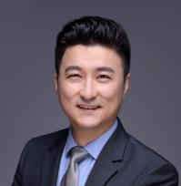
主讲人:卢虹冰 教授 空军军医大学
专家简介:
卢虹冰,空军军医大学生物医学工程系教授,主任,博士生导师,长期从事医学成像与智能分析研究,作为项目负责人近五年共承担国家重点研发专项、国家自然科学基金重点、国家科技支撑计划重大子课题、军队后勤科研重点项目等7项,2013年以第一完成人获陕西省科学技术一等奖1项;已在行业权威期刊Biomaterials, IEEE TMI, IEEE Trans Biomed Eng, ACS Appl Mater Interfaces, Euro Radiol等发表SCI论文70余篇,单篇最高引用近400次;获国家发明专利11项,美国专利1项,软件著作权5项;获 “国务院政府特殊津贴”、“军队优秀专业技术人才一类岗位津贴”等奖励。现任中国生物医学工程学会理事、中国图象图形学学会理事、陕西生物医学工程学会理事长等职,IEEE Trans Med Imag、《中国生物医学工程学报》等杂志编委。
授课内容:
医学图像质量评价方法对于提升医学图像质量、检验和优化医学图像处理算法等具有重要意义。主观评价方法存在代价高、耗时长、易受主客观因素影响,医学图像质量客观评价方法受到广泛关注。本讲座从临床案例出发,重点对基于检测和诊断任务的医学图像质量及分析算法的评价方法进行介绍,结合临床应用,对不同方法的作用及性能进行了分析。
课程题目5:Low-dose CT Quality Enhancement with Ethical AI
主讲人:Ge Wang Professor Rensselaer Polytechnic Institute
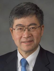
专家简介:
Ge Wang is the Clark & Crossan Endowed Chair Professor and Director of the Biomedical Imaging Center, Rensselaer Polytechnic Institute, Troy, NY, USA. He published the first spiral/helical cone-beam/multi-slice CT algorithm in 1991 and has since then systematically contributed over 100 papers to theory, algorithms, artifact reduction and biomedical applications in this important area of CT research. Currently, there are 100+ million medical CT scans yearly with a majority in the spiral/helical cone-beam/multi-slice mode. Dr. Wang’s group developed interior tomography theory and algorithms to solve the long-standing “interior problem” for high-fidelity local reconstruction, enabling omni-tomography (“all-in-one”) with CT-MRI as an example. He initiated the area of bioluminescence tomography with a significant impact on biophotonics. He has published 480+ journal publications, covering imaging-related diverse topics at depth and receiving a high number of citations and academic awards. His results were featured in Nature, Science, PNAS, and various news media. In 2016, he published the first perspective on deep-learning-based tomographic imaging as the new direction of machine learning and the Nature Machine Intelligence paper on the potential of deep learning over iterative reconstruction. His team has been continuously well-funded by federal agencies and major imaging companies, actively translating deep learning techniques into imaging products. His research interests include x-ray CT, MRI, optical tomography, multimodality fusion, and machine learning. He is the Lead Guest Editor for five IEEE Transactions on Medical Imaging Special Issues, Founding Editor-in-Chief of International Journal of Biomedical Imaging, Board Member of IEEE Access, and Associate Editor of IEEE Trans. Med. Imaging (TMI) (recognized as “Outstanding Associate Editor” by TMI), Med. Phys., and Machine Learning Sci. and Tech.. He is Fellow of IEEE, SPIE, OSA, AIMBE, AAPM, and AAAS.
授课内容:
Computer vision and image analysis are excellent examples of deep learning, which deal with existing images and produce features of these images (images to features), tomographic imaging produces images of multi-dimensional structures from experimentally measured features (integrals for CT) (features to images). Recently, deep learning is being actively developed for tomographic imaging, such as CT, forming a new area of imaging research. Currently, a hot debate continues on the pros and cons of AI-based imaging. Recently, the radiology societies in the United States, Europe, and Canada called for additional guidelines on the ethical use of AI. A joint statement was just published (https://pubs.rsna.org/doi/10.1148/radiol.2019191586) to “promote wellbeing, minimize harm, and ensure that the benefits and harms are distributed among the possible stakeholders in a just manner”. It is expressed that “radiologists will remain ultimately responsible for patient care in the new AI ecosystem”. We believe that the responsibility is upon not only radiologists but also PhD researchers. Preliminary results and considerations will be presented in this lecture, with an emphasis on low-dose CT imaging (https://www.nature.com/articles/s42256-019-0057-9).
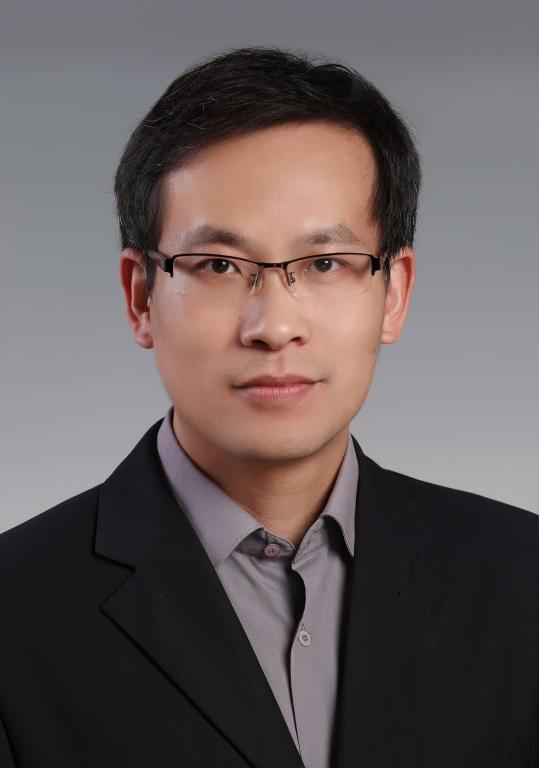
课程题目6:深度重建:基于深度学习的CT图像重建
主讲人:张意 副教授 四川大学
专家简介:
张意,四川大学计算机学院(软件学院)副教授,博士生导师,软件工程系系主任。从事医学物理以及图像反问题的相关工作,具体研究方向包括图像重建、稀疏表达、深度学习等。目前已在IEEE TMI、IEEE TCI、OE、BOE、JOSA A、Neurocomputing、European Radiology等权威国际学术期刊发表论文60余篇,多次受邀本领域多个主流会议(IEEE NSS/MIC, IEEE ISBI, Fully 3D,SIAM IS, SPIE Optics Engineering + Applications等)上作特邀报告,作为负责人主持国家重点研发计划子课题、国家自然科学基金(青年、面上),四川省科技支撑计划项目等多个国家级、省部级项目。中国生物医学工程学会医学图像信息与控制分会委员,中国自动化学会分数阶系统与控制专委会委员,中国计算机学会YOCSEF成都委员。IEEE Access副主编,International Journal of Biomedical Imaging客座编辑。
授课内容:
近年来,深度学习在计算机视觉、图像处理领域已经取得了丰硕的成果,但是在医学成像领域,主要的应用仍然停留在图像分析。基于深度学习的医学图像重建工作才刚刚起步,本报告主要介绍基于深度学习的图像重建领域的一些代表性工作成果和研究进展,同时将展望基于深度学习的图像重建方法未来发展的趋势。